Caas MR 4D Flow is specifically designed to extract relevant information from 3D phase-contrast (PC) MR images, within a few clicks. Based these phase-contrast images, two pioneering 4D Flow modules are offered to perform a complete flow analysis that can aid in reading and interpreting cardiovascular MR images and assist in planning the time and type of an intervention. These two modules include a module to analyze vessels, from the aorta up to smaller vessels in for example the kidneys (Caas MR 4D Artery), and a module to analyze the heart valves in terms of regurgitation fraction (Caas MR 4D Heart).
Key Results:
- Visualization of aortic blood flow as streamlines, pathlines and color-coded vectors
- Distribution of wall shear stress and local values of wall shear stress
- Pressure difference over a segment
- Visualization and quantification of regurgitant fraction over all four valves
- 2D flow phase-contract analysis
The Wall Shear Stress Analysis and Pressure Difference Analysis are not 510(k) cleared and therefore not meant for clinical decision making.
Key product features
- Intracardiac analysis with automated valve tracking
- Visualization and quantification of blood flow in just a few clicks
- Load and analyze 4D flow datasets from all main OEM vendors
Contact information
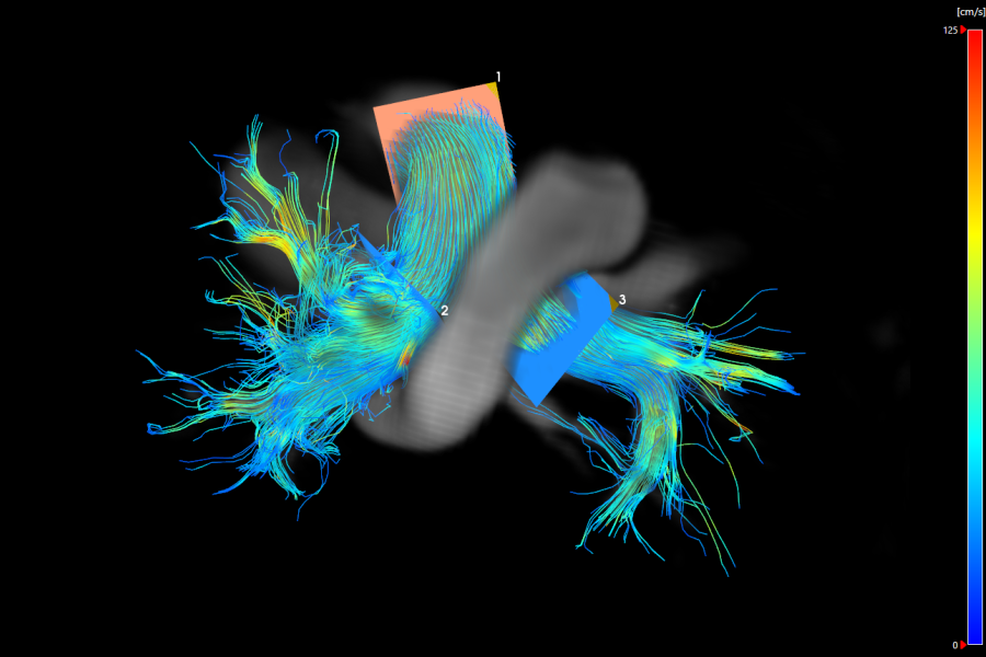
4D Artery - Streamlines in pulmonary arteries
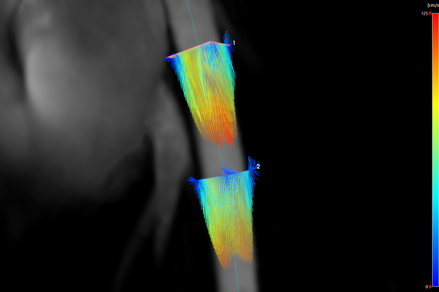
4D Artery - Vector Fields
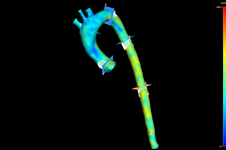
4D Artery - Wall Shear Stress
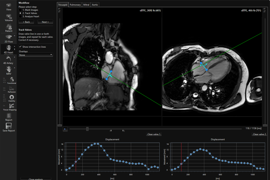
4D Heart - Automated Valve Tracking
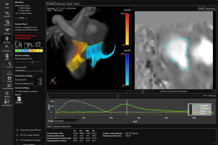
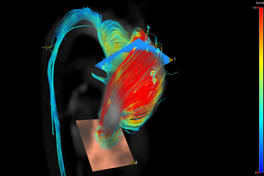
4D Heart - Streamlines over heart valves
- van der Palen, R.L.F., Roest, A.A.W. et al. Scan–rescan reproducibility of segmental aortic wall shear stress as assessed by phase-specific segmentation with 4D flow MRI in healthy volunteers. Magn Reson Mater Phy (2018). DOI: https://doi.org/10.1007/s10334-018-0688-6
- Bissell M. Phenotypes of aorta in AVD insights from imaging. Presented at EuroECHO Imaging 2016.
- Bissell M. 4D flow: Adding an extra dimension. Presented at EuroECHO Imaging 2016.
- Elders B et al. Ascending aortic wall shear stress and distensibility are different in patients with corrected atrioventricular septal defect compared to healthy controls: a comprehensive CMR and 4D flow MRI evaluation. JCRM 2016. 18(Suppl 1):P162
Why choose us?
We always aim to improve the quality and efficiency of cardiovascular image analysis to optimize patient treatment. To realize this, we:
- Provide fast and user-friendly software;
- Align our product portfolio with the latest developments in the cardiovascular field. In this way we aim to provide our customers with the optimal software solution at the right time;
- Offer training options which can be tailored to your needs.
We believe in the importance of training and support to assure that all our users are proficient and comfortable with their analysis. Please visit our Training & Support page for more information.