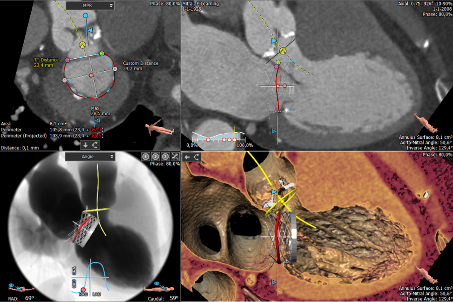
The Mitral Valve is a complex 3D structure. Mitral regurgitation can be treated by replacing the native valve (TMVR) or by repairing the native valve (TMVr). Planning for both types of procedures can be done using the 3mensio Mitral Valve workflow. With a single click we orientate on the mitral annulus. We can do an assisted mitral annulus trace to understand the 3D shape and dimensions of the annulus.
Additional features have been developed specifically to plan for replacement or repair. Relationships with surrounding structures like the Aortic Valve and Coronary Vessels can quickly be assessed. An easy comparison between the ED and ES phase can be performed. The approach route can be assessed using either the Transseptal or Direct Access approach module.
Key Results
General
- Dynamic visualization of mitral valve, left ventricle and left atrium
- Dimensions of Mitral Valve
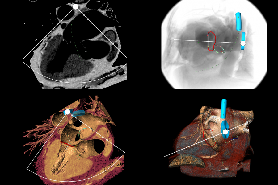
- Optimal C-arm angle
- Anatomical relations between interatrial septum, LAA ostium, mitral annulus and/or vena cava
- Volume of the left atrium or ventricle
TMVR
- Aortic-Mitral Angle
- Assessment of the neo-LVOT
- Device placement using Virtual Device
TMVr
- Distance annulus and coronary vessels
- Assessment of anchor positions
Key product features
- PACS connectivity
- Easily trace mitral annulus and evaluate the complexity
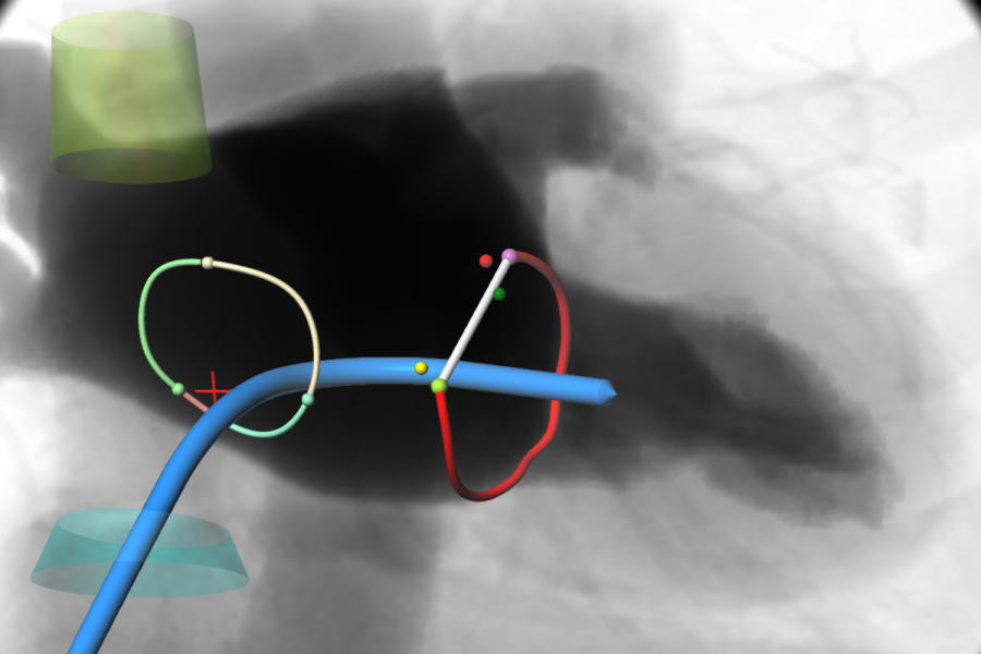
- Dedicated anchoring workflow (for repair methods)
- Transapical and Transseptal approach route modules
- .STL import
Contact information
Mitral Valve Workflow
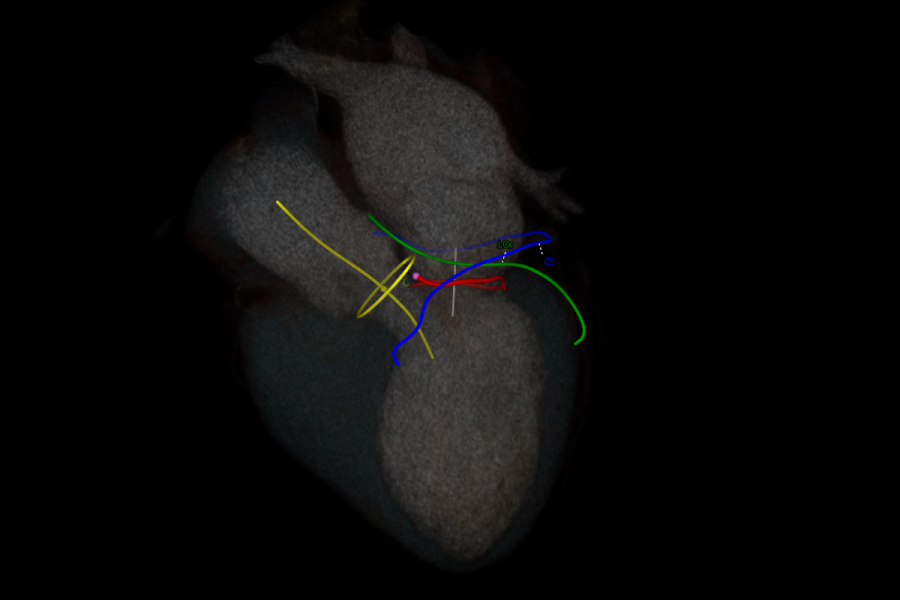
Overview Mitral and Aortic Valve and the CS and LCx
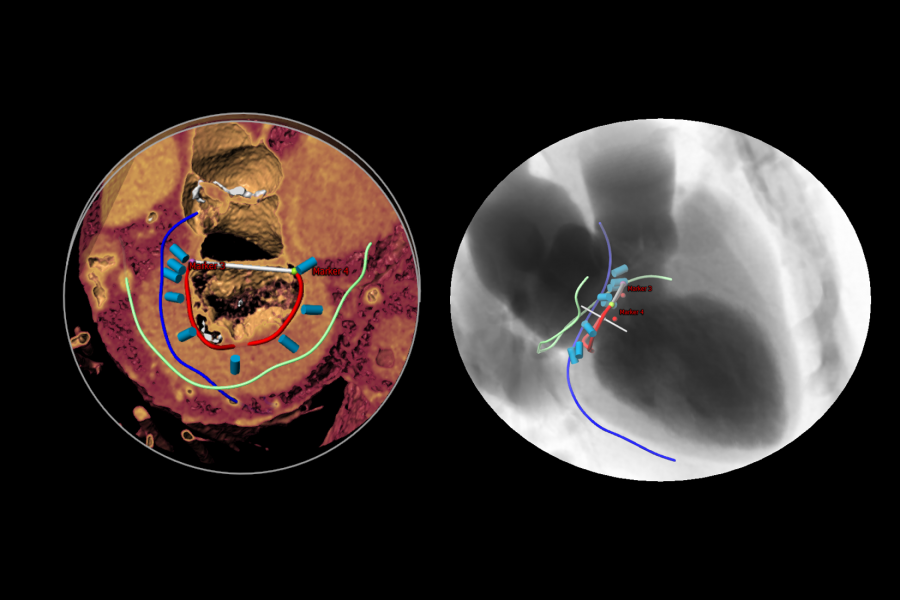
Mitral Anchoring Workflow
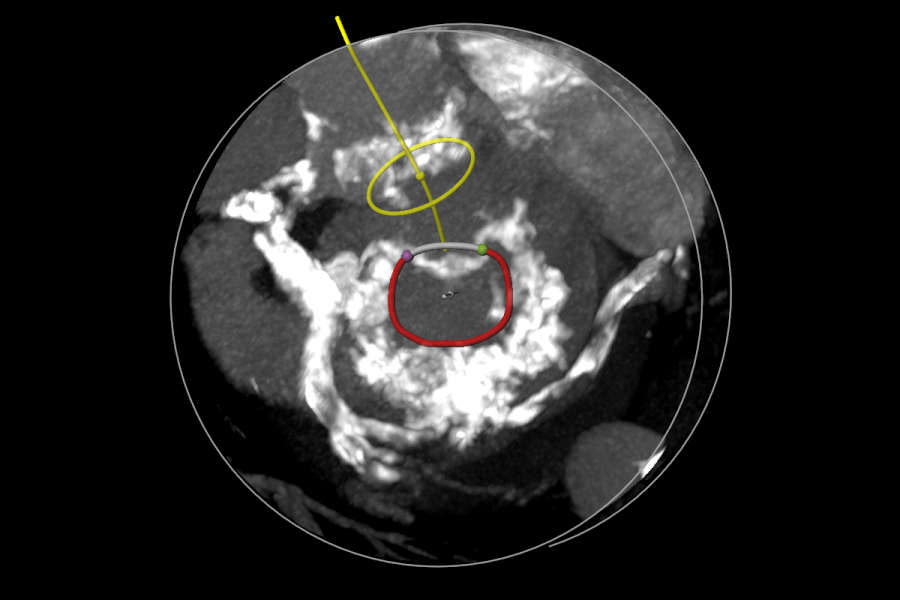
Mitral Annular Calcification
Septal Puncture Assessment
TEE Planning Simulation
- Abeldhani M et al. A simplified and reproducible method to size the mitral annulus: implications for transcatheter mitral valve replacement. European Heart Journal – Cardiovascular imaging 2016.
- Van Mieghem N, Rodríguez-Olivares R, et al. Computed tomography optimised fluoroscopy guidance for transcatheter mitral therapies. EuroIntervention 2016;11:1428-1431
- Piazza N et al. Quantitative multi-slice computed tomography assessment of the mitral valvular complex for transcatheter mitral valve interventions part 1: systematic measurement methodology and inter-observer variability. EuroIntervention 2015; 11.
- N. Piazza et al. Quantitative multi-slice computed tomography assessment of the mitral valvular complex for transcatheter mitral valve interventions part 2: geometrical measurements in patients with functional mitral regurgitation. EuroIntervention 2015; 11.
- Blanke P, Dvir D, Cheung A, et al. Mitral annular evaluation with CT in context of transcatheter mitral valve replacement. J Am Coll Cardiol Img. 2015; 8(5):612
- Blanke P, Dvir D, Naoum C et al. Prediction of fluoroscopic angulation and coronary sinus location by CT in the context of transcatheter mitral valve implantation. J Cardiovasc CT 2015; 9:183-192
- Gordic S, Nguyen-Kim TD, Manka R et al. Sizing the mitral annulus in healthy subjects and patients with mitral regurgitation: 2D versus 3D measurements from cardiac CT. Int J Cardiovasc Imaging 2014;30:389-98
- Alkadhi H, Desbiolles L, Stolzmann P et al. Mitral annular shape, size, and motion in normals and in patients with cardiomyopathy: evaluation with computed tomography. Invest Radiol 2009;44:218-25
Why choose us?
We always aim to improve the quality and efficiency of cardiovascular image analysis to optimize patient treatment. To realize this, we:
- Provide fast and user-friendly software;
- Align our product portfolio with the latest developments in the cardiovascular field. In this way we aim to provide our customers with the optimal software solution at the right time;
- Offer training options which can be tailored to your needs.
We believe in the importance of training and support to assure that all our users are proficient and comfortable with their analysis. Please visit our Training & Support page for more information.